Inhaltsverzeichnis
Why is bone grafting necessary?
The jawbone needs stimulation — without a tooth, it deteriorates. This principle explains why so many patients need bone grafting before implant placement. The bone that once supported a tooth is classified by the body as "unnecessary" without mechanical stimulation and is gradually resorbed — a process that begins just weeks after tooth loss.
The most common causes of jawbone loss are:
- Tooth loss: Within the first 12 months after extraction, the jawbone loses up to 25% of its volume. Bone resorption begins immediately and progresses continuously without intervention.
- Periodontitis: This chronic inflammation of the tooth-supporting structures destroys not only the gums but also the underlying jawbone — often insidiously and unnoticed for years.
- Long-term tooth loss: Those who wear removable dentures for years or decades steadily lose bone substance. The pressure from the denture further accelerates the deterioration.
- Accidents and trauma: Falls, sports injuries, or jaw fractures can cause acute loss of bone substance.
- Tumors and cysts: Benign or malignant changes in the jaw area can leave significant bone defects that must be rebuilt before implant placement.
Without treatment, progressive bone loss has far-reaching consequences: neighboring teeth lose their support and drift into the gap, the facial profile changes (sunken cheeks, wrinkle formation), and later implant placement becomes increasingly difficult — until at some point it becomes impossible without prior bone grafting.
The good news: With modern bone grafting procedures, we can reliably restore lost bone substance and thus create the foundation for permanent dental implants.
Overview of bone grafting methods
Depending on the extent and location of bone loss, various procedures are available. In our practice, we master the full spectrum of bone grafting surgery — from minimally invasive socket preservation to extensive bone block transplantation.
| Method | Description | Indication | Bone gain | Healing time |
|---|---|---|---|---|
| Bone augmentation | Bone substitute material is applied onto the alveolar ridge | Horizontal bone loss | Moderate (2–5 mm) | 4–6 months |
| Bone splitting / spreading | The alveolar ridge is split and expanded | Narrow alveolar ridge | Moderate (2–4 mm width) | 3–4 months |
| Bone block transplantation | Autogenous bone block is screwed in place | Large defect (vertical + horizontal) | Substantial (5–10 mm) | 4–6 months |
| Distraction osteogenesis | Bone is cut and slowly pulled apart | Vertical bone loss | Substantial (up to 15 mm) | 3–6 months |
| Socket preservation | Extraction socket is immediately filled | Immediately after tooth extraction | Volume preservation | 3–4 months |
Which method is right for you depends on your individual situation — the location and extent of the bone defect, general health, and planned implant restoration. During the consultation, we will discuss together based on CBCT 3D imaging which procedure promises the best outcome.
We frequently combine methods — for example, bone augmentation with membrane technology (GBR — Guided Bone Regeneration) to optimize results.
Sinus lift: Bone grafting in the upper jaw
The sinus lift is a specialized form of bone grafting developed specifically for the lateral upper jaw. In this area, the maxillary sinus lies directly above the tooth roots. After tooth loss, the sinus expands downward and the bone becomes too thin for implants.
During a sinus lift, the sinus membrane is elevated and the resulting cavity is filled with bone substitute material. Depending on the remaining bone height, two techniques are used: the internal sinus lift (minimally invasive, with 4–7 mm remaining bone height) and the external sinus lift (with less than 4 mm remaining bone height).
Since the sinus lift is a comprehensive topic in its own right, we have dedicated a separate page to it:
Treatment procedure
Bone grafting is a planned surgical procedure that requires careful preparation. In our practice, we follow a proven workflow that maximizes safety and precision:
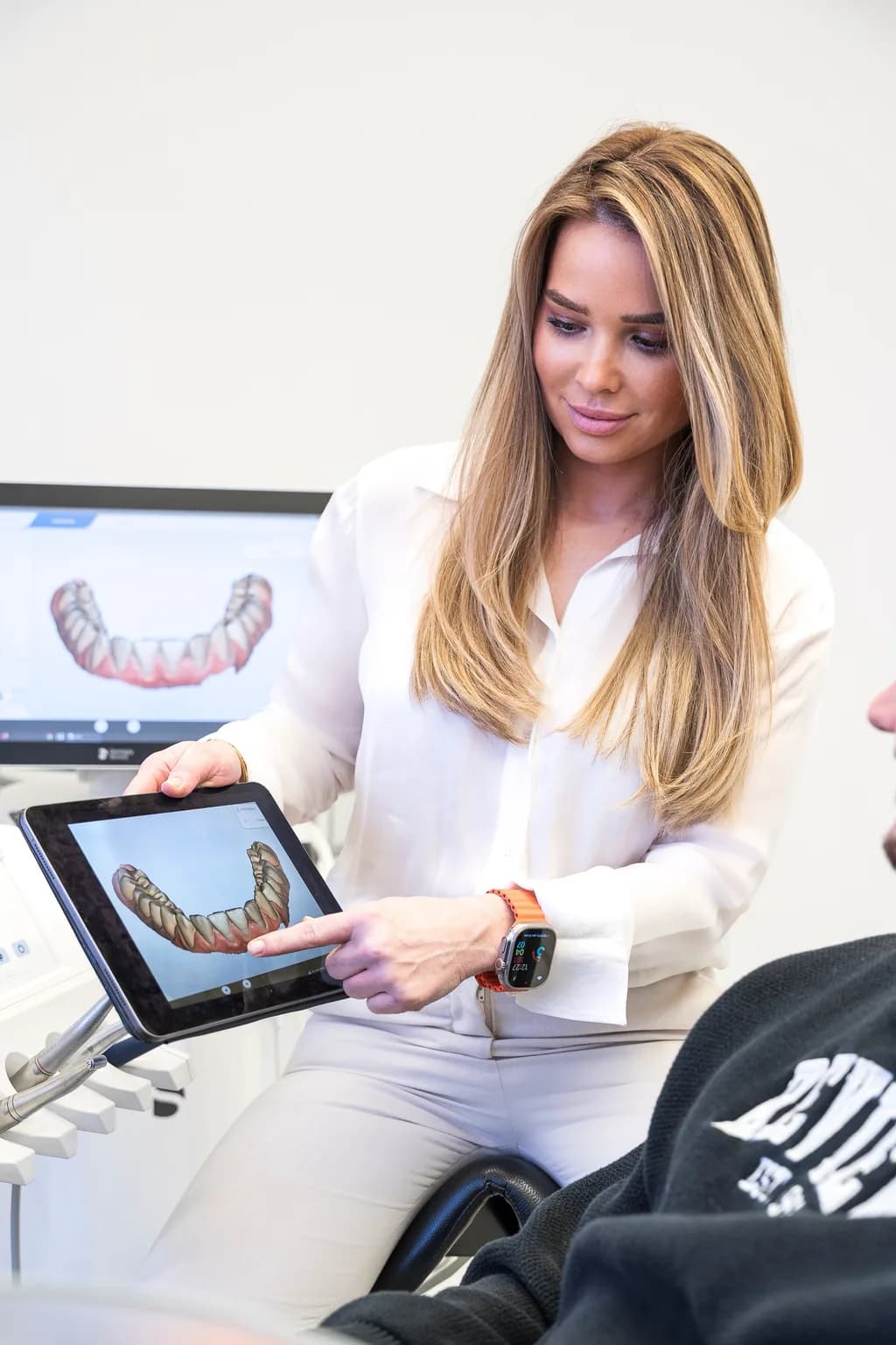
- CBCT 3D diagnostics: Using cone beam computed tomography (CBCT), we create a three-dimensional image of your jaw. This allows us to measure with millimeter accuracy how much bone is present, where nerves and sinuses are located, and how much bone needs to be built up.
- Treatment planning: Based on the 3D data, we create an individual treatment plan. We discuss with you the appropriate method, material, expected healing time, and the further course of treatment through to the finished implant.
- Material selection: Together, we decide whether autogenous bone, bone substitute material, or a combination will be used — depending on defect size, indication, and individual circumstances.
- Bone grafting surgery: The procedure is performed under local anesthesia. For anxious patients, we also offer nitrous oxide, twilight sedation, or general anesthesia. The duration varies depending on the method, between 30 minutes and 2 hours.
- Healing phase: Depending on the method, the grafted bone needs 3–6 months to remodel into solid, load-bearing bone. During this time, you wear a temporary prosthesis if needed.
- Implant placement: Once the bone has sufficiently healed — confirmed by a follow-up CBCT scan — we place the dental implant.
- Prosthetic restoration: After the implant has healed (an additional 3–6 months), the final restoration with a crown, bridge, or denture follows — your new, permanent dental prosthesis.
In certain cases, bone grafting and implant placement can be combined in a single session — the so-called simultaneous approach. Whether this is possible depends on the defect size and the primary stability of the implant.
Materials for bone grafting
Choosing the right bone grafting material is crucial for treatment success. In our practice, we use both autogenous bone and high-quality bone substitute materials — depending on the indication and individual situation.
Autogenous bone (autologous) — the gold standard
Autogenous bone has the highest biological value: it contains living bone cells, growth factors, and the body's own proteins that actively promote new bone formation. The bone is harvested from a suitable donor site — depending on the required amount, from the chin, the mandibular angle, or for larger defects from the iliac crest.
Advantage: Best healing, no rejection risk, contains bone-forming cells (osteogenic). Disadvantage: Secondary surgical site at the donor area, limited availability.
Xenograft material (animal-derived)
Bovine (cow-derived) bone substitute material such as Bio-Oss is one of the most extensively researched materials in implantology. All organic material is completely removed, leaving only the mineral bone structure — a safe, biocompatible scaffold into which the body can build new bone.
Advantage: No secondary surgical site, unlimited availability, well-researched, slow resorption supports the bone long-term. Disadvantage: Contains no living cells.
Alloplastic material (synthetic)
Synthetic materials such as tricalcium phosphate (TCP) or hydroxyapatite are manufactured in the laboratory and mimic the mineral structure of natural bone. They are fully resorbable — the body breaks them down over time and replaces them with its own bone.
Advantage: No secondary surgical site, no animal or human components, fully resorbable. Disadvantage: Slower new bone formation than with autogenous bone.
Allograft (human donor bone)
Human donor bone from a bone bank is rarely used in Germany but is available as an option. The material is extensively processed and sterilized to ensure maximum safety.
Combination of different materials
In practice, we frequently combine different materials to unite their advantages — for example, autogenous bone mixed with xenograft substitute material. This way, the patient benefits from the biological activity of autogenous bone and the volume of the substitute material. Additionally, we use collagen membranes (GBR technique) that protect the graft and guide new bone formation in a targeted manner.
Healing duration and aftercare
The healing time after bone grafting varies depending on the method used and individual healing progress:
- Socket preservation: 3–4 months
- Bone splitting / spreading: 3–4 months
- Bone augmentation: 4–6 months
- Bone block transplantation: 4–6 months
- Distraction osteogenesis: 3–6 months
- Sinus lift: 4–9 months (depending on internal/external technique)
Only after complete healing — verified by a follow-up CBCT scan — do we place the implant.
Aftercare in the first weeks:
- Soft diet: Only soft foods (soups, purees, yogurt) for the first 7–10 days. Avoid hard, crumbly, or spicy foods.
- Cooling: Apply cold compresses externally during the first 48 hours (20 minutes on, 20 minutes off) to minimize swelling.
- No smoking: Smoking is one of the greatest risk factors for complications. Abstain from nicotine for at least 2 weeks, ideally 8 weeks after the procedure.
- Gentle oral hygiene: Do not touch the wound area with a toothbrush for 1–2 weeks. Use an antiseptic mouthwash (chlorhexidine) instead.
- Follow-up appointments: Suture removal after 7–10 days. Further check-ups after 4 weeks and before the planned implant placement.
Special considerations for the upper jaw (after sinus lift or augmentation near the maxillary sinus):
- Do not blow your nose — only gently dab (2–3 weeks)
- Sneeze with your mouth open
- Do not fly for at least 2 weeks (pressure differences stress the maxillary sinus)
Costs and insurance coverage
The costs for bone grafting depend on the method, the extent of the defect, and the material used. Since every case is individual, we prepare a detailed cost estimate after the diagnostic examination.
Statutory health insurance (GKV)
Bone grafting is not covered by statutory insurance. The statutory health insurance funds cover neither the bone grafting nor the implant placement itself. What the GKV pays is the fixed subsidy for the final dental prosthesis — the crown, bridge, or denture that is attached to the implant. This fixed subsidy amounts to 60% of the standard care and can increase to up to 75% with a consistently maintained bonus booklet.
The bone grafting, bone substitute material, and implant placement are private services billed according to the German dental fee schedule (GOZ).
Private health insurance (PKV)
Private health insurance companies reimburse bone grafting procedures partially or fully under many plans — particularly when there is a medical necessity for the subsequent implant placement. We recommend submitting the cost estimate to your private insurance before starting treatment. We are happy to assist you with this.
Installment payment
To ensure that costs do not stand in the way of optimal care, we offer flexible installment payment plans. This allows you to spread the costs for bone grafting and implant placement over several months. Please feel free to ask us about this during the consultation.
Why choose Dr. Dickel for your bone grafting?
Bone grafting is one of the most demanding disciplines in dentistry. Experience, modern diagnostics, and surgical skill are crucial for a safe and long-term successful outcome. In our practice in Munich-Oberfoehring, we combine all the prerequisites for excellent bone grafting surgery:
- CBCT 3D diagnostics: Millimeter-precise three-dimensional imaging for accurate planning — no guesswork, but exact measurement of available bone.
- DGI member: Member of the German Society for Implantology — the leading professional society for implantology in Europe.
- DGZMK and DGAEZ: Member of the German Society for Dental, Oral and Maxillofacial Medicine and the German Society for Aesthetic Dentistry.
- CEREC technology: Digital CAD/CAM fabrication of dental prosthetics in a single session — for fast and precise prosthetic restoration after bone grafting.
- Full range of methods: We master all established bone grafting procedures — bone augmentation, bone splitting, bone block transplantation, distraction osteogenesis, socket preservation, and sinus lift. This way, we find the optimal solution for every defect.
- All sedation options: From nitrous oxide to twilight sedation to general anesthesia — we offer anxious patients and extensive procedures the appropriate sedation.
- Autogenous bone and bone substitute material: We work with both material groups and combine them individually for the best result.
Schedule a consultation — we will analyze your situation with state-of-the-art 3D diagnostics and create an individual treatment plan for your bone grafting.
Kosten im Überblick
| Leistung | Preisrahmen | Hinweis |
|---|---|---|
| Simple bone grafting (augmentation) | Private service per GOZ | Bone augmentation or bone splitting for moderate defects. Costs vary depending on material and extent. Not covered by statutory health insurance. |
| Extensive bone grafting / bone block | Private service per GOZ | Bone block transplantation or distraction osteogenesis for larger defects. Higher effort due to donor site and longer surgery time. |
| Socket preservation | Private service per GOZ | Filling of the extraction socket immediately after tooth removal. Comparatively low effort to preserve bone volume. |
Simple bone grafting (augmentation)
Private service per GOZ
Bone augmentation or bone splitting for moderate defects. Costs vary depending on material and extent. Not covered by statutory health insurance.
Extensive bone grafting / bone block
Private service per GOZ
Bone block transplantation or distraction osteogenesis for larger defects. Higher effort due to donor site and longer surgery time.
Socket preservation
Private service per GOZ
Filling of the extraction socket immediately after tooth removal. Comparatively low effort to preserve bone volume.
Private health insurance companies frequently reimburse bone grafting procedures partially or fully, provided there is a medical necessity for the implant placement. We are happy to prepare a detailed cost estimate for your private insurance.
For bone grafting and implant placement, we offer flexible installment payment plans so that costs do not stand in the way of your optimal care.
Risiken und Sicherheit
Bone grafting procedures are routine operations in dental surgery today and are very safe when carefully planned and executed. However, as with any surgical procedure, complications can occur.
Swelling and bruising
Common (normal)
Normal reaction to the procedure. Cooling during the first 48 hours, swelling subsides within 5–7 days.
Post-operative bleeding
Occasional
Slight bleeding is normal in the first hours. For heavier bleeding: use a gauze pad and contact the practice.
Infection
Rare
Prophylactic antibiotics, sterile operating conditions, and careful aftercare minimize the risk.
Graft failure / poor integration
Rare
Consistently avoid risk factors such as smoking and poor oral hygiene. If integration fails, a repeat bone grafting is possible.
Nerve damage in the lower jaw (inferior alveolar nerve)
Rare
CBCT 3D planning enables precise visualization of the nerve pathway. A safety margin is maintained. Numbness is usually temporary.
Rejection of foreign material
Very rare
Bone substitute materials are highly biocompatible. In case of intolerance, the material is removed and an alternative is chosen.
In our practice, we minimize risks through CBCT 3D planning with millimeter-precise visualization of nerves and bone structures, sterile operating conditions, minimally invasive techniques where possible, close follow-up care, and individual counseling on risk factors (smoking, medications, pre-existing conditions).
Qualifikationen und Zertifikate
DGI — German Society for Implantology
Membership
DGZMK — German Society for Dental, Oral and Maxillofacial Medicine
Membership
DGAEZ — German Society for Aesthetic Dentistry
Membership
CEREC Certification — Digital CAD/CAM dental prosthetic fabrication
Certified
CBCT 3D Diagnostics — Cone Beam Computed Tomography
Practice equipment
2012
State examination in dentistry
University
Häufige Fragen
Ready to take your first step?
Book an appointment for a personal consultation at our practice in Munich Oberföhring.